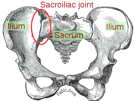
Form Closureは、骨盤の解剖学的設計から関節の安定性を説明するものである。 仙骨と腸骨はそれぞれ1つの平らな面と1つの隆起した面を持ち、それらがかみ合うことで安定性が促進される。 左右対称の溝と隆起は、二関節関節の中で最も高い摩擦係数を実現し、関節を剪断から保護します。 SIJの骨の位置は、骨盤の輪の安定性を高める「楔石」のような形状を作り出しています。 仙骨は上側が広くなっているため、仙骨が腸骨の間に「挟まる」ことができ、この「楔石」形状が作られるのである。
Force Closure
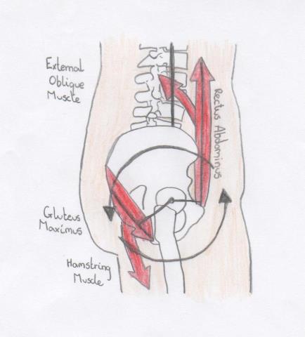
form closureはSIJに安定性を与えるが、可動性が生じるためには、垂直荷重に耐えられるようにさらに関節を圧迫して安定させることが必要である。 フォースクロージャーとは、安定性を生み出すために関節全体に作用する他の力を表す言葉である。 この力は、仙腸関節に垂直な繊維方向を持つ構造物によって発生し、負荷の状況に応じて調節可能です。 筋肉、靭帯、胸腰部フェイシアのすべてがフォースクロージャーに寄与しています。
Forst S.L, Wheeler M, Fortin J.D and Vilensky J.A. Sacroiliac Joint(仙腸関節). 仙腸関節:解剖学、生理学および臨床的意義. ペインフィジシャン。 2006;9:61-68
3.0 3.1 3.2 3.3 3.4 3.5 Cohen S.P. Sacroiliac Joint Pain: A Comprehensive Review of Anatomy, Diagnosis, and Treatment. Anesthesia & Analgesia 2005: 101:1440-53
4.0 4.1 4.2 Arumugam A, Milosavljevic S, Woodley S and Sole G. Effects of external pelvic compression on form closure, force closure, and neuromotor control of the lumbopelvic spine.The Effect of external pelvic pressure on the future. システマティックレビュー。 Manual Therapy 2012; 17: 275-284
Mitchell T.D, Urli K.E, Breitenbach J & Yelverton C. The predictive value of the sacral base pressure test in detecting specific types of sacroiliac dysfunction(仙腸関節機能障害の特定タイプの検出における仙骨底圧テストの予測価値). カイロプラクティック医学ジャーナル 2007: 6, 45-55
6.0 6.1 6.2 6.3 Vleeming A, Stoeckart R, Volkers, ACW, Snijders CJ.仙骨底圧検査。 仙腸関節の形態と機能の関係。 Part 1: 臨床解剖学的側面。 Spine 1990a; 15(2): 130-132
SI Bone. SI関節の解剖学、バイオメカニクスと有病率。 Available from: http://www.youtube.com/watch?v=D6NTMgWCSaU
8.0 8.1 8.2 8.3 8.4 8.5 8.6 8.7 8.8 Willard F.H, Vleeming A, Schuenke M.D, Danneels L & Schleip R. the thoracolumbar fascia: anatomy, function and clinical considerations. 2012; 221(6): 507-36
9.0 9.1 9.2 9.3 9.4 9.5 9.6 9.7 9.8 Pool-Goudzwaard A.L, Vleeming A, Stoeckart R, Snijders C. J & Mens J.M.A. Insufficient lumbopelvic stability: a specific’ low back painへの clinical、解剖学、生体力学からのアプローチ. マニュアルセラピー。 1998; 3(1): 12-20
10.0 10.1 10.2 10.3 Liebenson C. The relationship of the sacroiliac joint, stabilization musculature, and lumbo -pelvic instability. ジャーナル・オブ・ボディワーク・アンド・ムーブメント・セラピーズ 2004:8:43-45.
12.0 12.1 Harrison DE, Harrison DD & Troyanovich SJ. 仙腸関節:解剖学とバイオメカニクスのレビューと臨床的意味合い。 仙腸関節:解剖学と生体力学のレビューと臨床的意義.
13.0 13.1 13.2 Van Wingerden JP, Vleeming A, Buryuk HM, Raissadat. 仙腸関節の生体内での安定化:骨盤の力閉鎖に対する筋の寄与の検証。 ヨーロッパスパインジャーナル 2004.
Vleeming A, Volkers ACW, Snijder CJ, Stoeckart R. Relation between form and function in the sacroiliac joint. 第2部:バイオメカニクス的側面。 また、仙腸関節の形態と機能との関係、第2部:生体力学的側面、Spine 1990b; 15(2):133-136
Snijder CJ, Vleeming A, Stoeckart R. 腰仙骨荷重の腸骨および脚への移動。 第1部:仙腸関節のセルフブレーシングのバイオメカニクスとその治療および運動における意義. 仙腸関節のセルフブレーシングのバイオメカニクスと治療と運動の意義. NZ Journal of Physiotherapy 2005 33:(3); 91-94
17.0 17.1 17.2 17.3 17.4 Vleeming A, De Vries H.J, Mens J.M.A & Van Wingerden J.P. Potossible role of the long dorsal sacroiliac ligament in women with peripartum pelvic pain. 仙結節と仙棘靭帯-仮想再建-. Ann Anat 2009: 191; 417-425.
Bechtel R. Human sacroiliac jointの軸索間靭帯の物理的特性。
Pool-Goudzwaard A, HoekvanDijke G, Mulder P, Spoor C, Snijders C & Stoeckart R. 腸腰椎靭帯:仙腸関節の安定性に対するその影響について. このような場合、仙腸関節の安定性に影響を与える。 仙腸関節:その解剖学的構造、機能および潜在的な臨床的意義の概要。 Journal of Anatomy 2012:221:6:537-67
22.0 22.1 Pel JJM, Spoor CW, Pool-Goudzwaard AL, Hoek van Dijke GA, Snijers CJ. 骨盤の筋肉と靭帯の力の最適化による仙腸関節のせん断荷重の軽減に関するバイオメカニクス的分析。 Ann Biomed Eng 2008; 36:3: 415-424
23.0 23.1 Richardson CA, Snijders CJ, Hides JA, Damen L, Pas MS, Storm J. The Relationship Between the Transversus Abdominis Muscles, Sacroiliac Joint Mechanics, and Low Back Pain. Spine 2002:27:4:399-405.
24.0 24.1 Ireland ML, Ott SM. 妊娠が筋骨格系に及ぼす影響. CLINICAL ORTHOPAEDICS AND RELATED RESEARCH 2000;372:169-179
28.0 28.1 Mantle J, Haslam J, Barton S. Physiotherapy in Obstetrics and Gynaecology(産科と婦人科における理学療法). 第2版。 ロンドン。 Elseiver Limited, 2004.
29.0 29.1 Mitra R. Osteitis Condensans Ilii. Rheumatology International 2009;30:293-296
Nicholas G. Demy osteitis condensans ilii.(ニコラス・デーミー・コンデンサンス・イリー骨炎). Lancet 1975;305(7916): 1135-1136
Hare HF, Haggart GF. 腸管凝縮性骨炎。 米国医師会雑誌 1945;128:723-727
Ritchie JR. 妊娠中の整形外科的考察。 また、「妊娠に関連する骨盤帯の痛みと妊娠中のリラキシン濃度との関係:システマティックレビュー」。 European Spine Journal 2012;21:1769-1776
34.0 34.1 34.2 34.3 34.4 34.5 34.6 Gilleard W, Brown M, Structure and Function of the Abdominal Muscles in Primigravid Subjects during Pregnancy and the Immediate post-birth period. Physical Therapy 1996;76(7):750-762
35.0 35.1 DiFiore, F. The Complete Guide to Postnatal Fitness(産後フィットネス完全ガイド). 第3版。 ロンドン。 A and C Black Publishers Ltd. 2010. p27.
36.0 36.1 Ricci S, Kyle T. Maternity and Paediatric Nursing. Philadelphia: をご覧ください。 2009.
Viktrup L, Lose G, Lower urinary tract Symptoms 5 years after the first delivery.は、最初の出産から5年後の下部尿路症状。 Int Urogynecol J 2000;11: pp 336-340。
Pairman S, Tracy S, Thorogood C, Pincombe J. Midwifery preparation for practise.日本助産学会雑誌. 第2版. Chatswood: チャーチルリビングストンエルゼビア。 2010. p407.
MacLennan A, Taylor A, Wilson D, Wilson D. The prevalence of pelvic floor disorders and their relationship to gender, age, parity and mode of delivery.骨盤底部障害の有病率と性別、年齢、分娩様式との関係.骨盤底部障害の有病率と性別、年齢、分娩様式との関係. 産科婦人科の英国ジャーナル2000;107:1460から1470まで。
Lee D. The Pelvic Girdle: An integration of clinical expertise and research. Forth Edition。 Edinburgh: チャーチルリビングストン。 2011.
Kassai K, Perelli K. The bathroom Key: 失禁に終止符を打つ。 ニューヨーク。 バン印刷。 2012.
50.0 50.1 Pool-Goudzwaard A, Slieker ten Hove M, Viethout M, Mulder P, Pool J, Snijders C, Stoeckart R. Relationship between pregnancy-related lower back pain, pelvic floor activity and pelvic floor dysfunction. 国際泌尿器科ジャーナル 2005;16: pp 468-474.
MacLennan A, Taylor A, Wilson D, Wilson D. The prevalence of pelvic floor disorders and their relationship to gender, age, parity and mode of delivery. 産科婦人科の英国ジャーナル2000;107:1460から1470まで。
Lal M, Mann H, Callender R, Radley S. 帝王切開分娩は肛門失禁を防ぐか? 産科と婦人科2003;101(2):305から312まで。
Sjodah J. 妊娠に関連した骨盤帯の痛みとその筋機能との関連. Linkoping: 大学. Linkoping. 2010.
Stuge B, Laerum E, Kirkesola G, Vollestad N. The Efficacy of a Treatment Program Focusing on Specific Stabilizing Exercises for Pelvic Girdle Pain After Pregnancy.(「妊娠後の骨盤帯の痛みに対する特定の安定化エクササイズに焦点を当てた治療プログラムの効果」): 無作為化対照試験。 Spine 2004;29(4):351-359